Delving deeper into organ development requires long-term monitoring of organ growth. INRAE researchers thus designed a novel approach that they employed to characterise growing leaves. Over several weeks and using many plants, the scientists photographed more than 1,000 leaves at varying developmental stages. By applying targeted algorithms to the photographs, they were able to reconstruct the complete story of leaf development. Published in Quantitative Plant Biology, their results highlight specific key steps in leaf growth, knowledge that could guide future strategies for optimising plant growth in general. Their unique approach is easy to use and may be applied to all types of organs in plants and animals alike.
In living organisms, development is a combination of multiple coordinated processes that interact in time and space over the course of growth. One false note in the delicate symphony can have catastrophic consequences. However, the precise score of this biological music is often a mystery to scientists. One solution is to use advanced microscopy techniques to observe organ growth in real time. The challenge is that such approaches are poorly suited to long-term monitoring. Furthermore, they can stress organs, disrupting development.
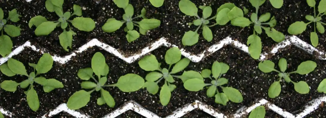
To address this issue, INRAE researchers developed a unique method that they have applied to plants. First, drawing upon numerous individual plants, scientists photograph leaves representing the range of developmental stages. Applying targeted algorithms to this photo catalogue, it is possible to assign an age to each leaf and reconstruct development over time. To help them in this task, the researchers exploit an open-source software programme—MorphoLeaf—which they developed in 2016 in collaboration with ENS de Lyon. The next step was to use the method to comprehensively document leaf development in the model plant Arabidopsis thaliana. Over a period of several weeks, scientists collected leaves from hundreds of wild-type and mutant strains; the latter had genetic modifications affecting leaf shape. A major advantage of this method is its ease of implementation: it requires no sophisticated equipment. Indeed, a standard microscope was used to take pictures of the earlier developmental stages (leaf size < 0.1 mm), and a flatbed scanner was used for the later developmental stages (leaf size > 1 cm).
The images told a precise story of leaf development, clarifying key points in space and time that contribute greatly to development in general. Notably, the analysis revealed that wild-type and mutant plants may have identical early developmental trajectories that then diverge at specific later stages. This detailed understanding of plant development can inform future strategies for optimising plant growth. The researchers’ approach is extremely flexible and can be adapted to any organ type, including in animals. It could thus be a valuable tool in studies seeking to demystify organ and organismal development.
Read the paper: Quantitative Plant Biology
Article source: INRAE
Image: Model plant Arabidopsis thaliana. Credit: INRAE – Jubault
